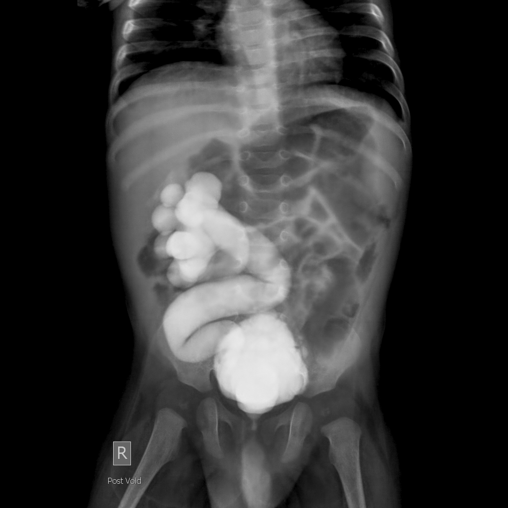
47+ Posterior Urethral Valve X Ray Images. What causes posterior urethral valves? At present, the diagnosis of posterior urethral valves is prenatal and the pattern is characterized by detrusor hypertrophy and more or less marked hydroureteronephrosis.
The images will show if there is any reverse flow of urine into the ureters and kidneys.
This valve is the most common cause of bladder outlet obstruction in male children. The cystoscopy however revealed a single. Antenatal and postnatal ultrasound (during and after your. Posterior urethral valves (puv) are obstructive membranes that develop in the urethra (tube that drains urine from the bladder), close to the bladder.
Comments
Post a Comment